

Understanding Facts about Brain Cancer
A brain tumour is a life-changing diagnosis that can affect both a person and their loved ones. These tumours arise from brain tissue or surrounding structures such as the meninges.
Primary brain cancers develop within the skull, whereas secondary brain cancers spread from other organs such as the lungs, liver, colon, breast or bone. Secondary tumours are far more common. While primary tumours may be benign or malignant, secondary brain tumours are always cancerous.
Among primary tumours, gliomas are the ones most likely to be malignant. Meningiomas tend to be benign. Within the glioma group, glioblastoma (grade IV glioma) is known for being particularly aggressive.
Common Symptoms of Brain Tumours

Man Experiencing Severe Headache Pain
Symptoms vary widely because they depend on the tumour’s location within the central nervous system. Common warning signs include:
-
Persistent headaches
-
Visual changes
-
Epileptic seizures
-
Loss of consciousness
-
Difficulty walking or coordinating movements
-
Weakness or paralysis in parts of the body
-
Mood or behavioural changes
How Tumour Location Affects Symptoms
Frontal lobe tumours may cause mood, behaviour and speech disturbances.
Tumours in the brainstem — located at the back of the brain — can interfere with breathing, swallowing and limb strength. Severe cases may lead to unconsciousness.
Tumours in the cortex commonly trigger epileptic seizures.
Tumours on the dominant side of the brain often present differently from those on the non-dominant side. The speed at which a tumour grows also influences symptoms. Slow-growing tumours may cause subtle, gradually progressive issues, while fast-growing tumours often cause sudden or severe symptoms.
Although headaches are usually caused by benign conditions, headaches from tumours in the lower or back part of the brain can be intense, may come with projectile vomiting, and typically do not respond to over-the-counter painkillers. Raised intracranial pressure may also be seen during a detailed eye examination. Sudden bleeding inside a tumour can also cause rapid worsening of symptoms.
Hormone-Producing Tumours
Pituitary tumours — usually benign — can cause hormonal changes.
Examples
-
Prolactinomas (microadenomas)
Increase prolactin levels, sometimes causing unexpected milk discharge. -
Somatotroph adenomas (macroadenomas)
Produce excess growth hormone, leading to gigantism or acromegaly.
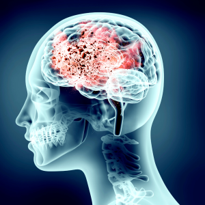
Detecting Brain Tumours

x-ray of brain showing tumor
Brain tumours are commonly identified through imaging.
Imaging Techniques
-
CT scans
-
MRI scans, often with contrast dye to help differentiate tumour types
-
PET scans, used in select cases to assess cancer spread
Other Diagnostic Tools
-
Comprehensive eye examinations
-
MR spectroscopy
-
Brain biopsy for definitive diagnosis
-
Blood tests and tumour markers (useful mainly for identifying secondary tumours)
Many tumours are detected early through routine health checks, which often leads to better outcomes.
Risk Factors and Prevention
The exact cause of most brain tumours remains unknown. However, linked risk factors include:
-
Advancing age
-
Male sex
-
Non-Hispanic white ethnicity
-
Exposure to chemicals
-
Exposure to ionising radiation
Research into war-zone exposures also suggests that certain chemical agents may elevate the long-term risk of brain cancer.
How Brain Tumours Are Treated
Treatment depends on factors such as:
-
Whether the tumour is cancerous
-
Its grade
-
Its size
-
Its location in the brain
Options include:
-
Observation
-
Surgical removal
-
Radiosurgery
-
Chemotherapy
Often, surgery is combined with radiotherapy or chemotherapy to achieve better outcomes.
Examples to Note
-
WHO Grade I glioma (pilocytic astrocytoma)
Often benign; surgical removal alone is usually sufficient. -
High-grade gliomas (e.g., glioblastoma)
Carry a higher risk of recurrence and require multimodal treatment.
Complete surgical removal of the tumour with clean margins offers the best chance of preventing recurrence. Tissue samples from surgery guide decisions about radiotherapy or chemotherapy. Many people receive oral chemotherapy drugs when needed.
Keeping an Eye Out: When to Seek Medical Attention

Doctor with human brain anatomy model.
Early detection improves the chances of successful treatment. Seek medical advice if you notice:
-
Sudden behavioural changes
-
A severe, unexplained new headache
-
Progressive weakness in any body part
-
New visual symptoms
-
Unexplained milk secretion
-
Excessive body growth
-
Difficulty speaking or performing fine hand movements
-
Difficulty walking
-
New-onset seizures
-
Urinary incontinence or poor balance
Even if these symptoms turn out to be benign, it is always safer to obtain a professional evaluation. PRIME


Leave A Comment