


Common Non-invasive Cardiac Tests and What They Mean
There are many cardiac tests which are commonly done as part of health screening. It is important to know what they mean so that we may know the appropriate steps to take following their results. Let us take a look at how we determine someone’s cardiovascular risk, the recommended cardiac tests to take and how to interpret your results.
Predicting Cardiovascular Risk
Cardiovascular risk assessment is the assessment of the risk of heart attack or stroke occurring in the next 5 to 10 years. We can predict cardiovascular risk based on gender, age and presence of risk factors, such as hypertension, diabetes, hyperlipidaemia and smoking.
In general, age and gender correlate to increased cardiovascular risk: older individuals and males are more likely to have cardiovascular disease compared to the young and females. Having a family history of a young family member suffering from coronary artery disease also increases the risk of heart attack and stroke.
Tests to Assess Cardiovascular Risk
HsCRP is a blood test which can be used as a supplement to assess general cardiovascular risk. If it is elevated, the likelihood of stroke and heart attack may be higher. However, it is not the sole marker of cardiovascular risk. In a clinical setting, this marker is usually assessed in conjunction with cholesterol, blood pressure, presence of diabetes, and whether the patient smokes.

There are several scoring systems which take into account all these cardiovascular risk factors and compute them on the basis of available scientific evidence and literature. Some of them include the Pooled Cohort Equations and the Qrisk3 score. These scoring systems can be easily found on Google and they can be conveniently used by lay persons. By keying in specific variables on these platforms, these platforms would provide us with a cardiovascular risk assessment.
Although different scoring systems may give slightly different numbers, we can generally get a ballpark cardiovascular risk from these publicly assessable scoring systems.
Requirements for Cholesterol and Diabetic Screening
While there may be a common perception that fasting is necessary before giving blood samples for cholesterol and diabetic health screening, the fact is that we now do not need fasting blood samples for cholesterol and diabetic screening.
Traditionally, blood tests for cholesterol and sugars need to be done on fasting blood samples. Patients need to fast for at least 10 hours prior to the samples being drawn. This is because cholesterol measures are calculated based on triglycerides (blood fat) in the blood which may be affected by food intake.
However, laboratories today can measure bad LDL (low-density lipoproteins) cholesterol levels directly without the need for fasting. Blood sugar measurements may be measured in a non-fasting sample too. HbA1c, which is a reflection of the average blood sugar in the preceding 3 months, does not require fasting either.
Other Tests for Coronary Evaluation
There are several ways to evaluate for coronary artery disease. We can either evaluate the arteries directly with a coronary angiogram or with a CT-coronary angiogram. They allow direct visualisation of the arteries by detecting contrasts in the coronary arteries. A CT-coronary angiogram would also provide us with a calcium score which is a reflection of the overall health of one’s coronary arteries. A higher calcium score means that there is more calcium being deposited on the coronaries and that would translate to poorer overall cardiovascular health outcomes.
We also have stress tests which actually assess the ability of the coronary arteries to supply adequate blood flow to the heart. The concept is that if there is stenosis of the coronary arteries, the perfusion to the myocardium may be affected during states of stress even when it is normal at rest.
Although a stenosed artery can provide adequate blood flow at rest, when the demand for blood becomes higher at stress, a stenosed artery may fail to provide adequate blood flow to the tissues at stress.
Stress Tests
Today, we have myocardial perfusion scans which measure how well the myocardium is perfused by blood. By injecting radioactive isotopes in the blood, the blood flow within the heart muscles can be visualised using a special scanner which can detect the small amounts of radiation emitted by these isotopes.
The assessment of perfusion is done twice — once at rest and once at stress. This is because a stenosed coronary artery may still be able to supply adequate blood to the myocardium in a resting state but not in a stressed state. Hence, for a patient with stenosis of a coronary artery, we would expect normal perfusion at rest and impaired perfusion at stress.
A stress echocardiogram utilises a similar premise. It is done at rest and under stress, and it evaluates the heart muscle directly. If perfusion to a specific part of the heart muscle is affected, it may not contract as well in a stressed state, thus allowing perfusion to be assessed indirectly.
One stress test that is readily available is the treadmill ECG. A treadmill ECG is done by asking a patient to run on the treadmill while monitoring his/her blood pressure, heart rate and ECG. Several useful information can be obtained from a treadmill ECG which may include exercise tolerance, cardiac symptoms and ECG changes during exercise.
Stressing the Heart during Stress Tests
There are several ways to stress the heart. The best way would be exercise since we can obtain information about cardiovascular fitness as well, which can predict cardiovascular risk.
In stress tests, exercise can be done either on a treadmill or on a stationary bike. A stationary bike may be well suited for patients who have mobility limitations, such as osteoarthritis of the knees.
For myocardial perfusion scans and stress echocardiograms, stress can also be induced by injecting medications directly into the veins. This is done when patients are completely unable to exercise. While some of these medications increase the heart rate, other medications dilate the heart arteries directly.
Normal vs. Positive Treadmill ECG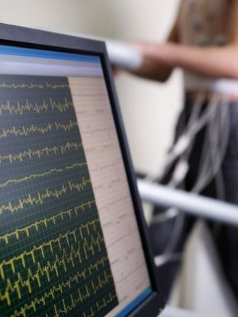
The interpretation of a normal or positive treadmill ECG can be very tricky as it really depends on the patient’s pre-test likelihood of coronary artery disease. Furthermore, a treadmill ECG also carries limitations in sensitivity and specificity. As such, although a treadmill ECG is often used to suggest absence or presence of disease, we need to interpret the results in the context of the patient and the limitations of the ECG.
If the treadmill ECG is normal in a patient with a high risk of coronary artery disease, there is still a high chance he has significant coronary artery disease. An analogy may be the forecasting of sunny weather with no rain despite the presence of dark clouds in the sky. We are likely to take such a weather forecast with a pinch of salt. In the same vein, we will also take a normal treadmill ECG in a patient whom we feel is highly likely to have coronary artery disease (based on clinical assessment) with a pinch of salt.
On the other hand, if a treadmill ECG is positive in a patient whom we feel have very low risk of coronary artery disease, we need to think twice too. Like the earlier analogy, this reading is akin to getting a weather forecast of massive thunderstorms half an hour later when we are seeing clear sunny skies right now.
In such situations, the patients may need further in-depth coronary evaluation with imaging, such as a CT-coronary angiogram, to further clarify or confirm the diagnosis.
We often depend on weather forecasts, instead of relying on our gut feel, to plan our daily activities. Likewise, cardiac tests like the treadmill ECG help us to predict the presence of coronary artery disease in patients with moderate cardiovascular risk.
A normal treadmill ECG result in a patient with a moderate likelihood of having coronary artery disease may reassure us. On the other hand, a positive treadmill ECG in a patient with moderate likelihood of having coronary artery disease may warrant more in depth coronary evaluation and aggressive cardiovascular risk management.
Key Takeaways
In general, when a non-invasive test in normal in an individual, the risk of a heart attack or stroke occurring in the next 5 years is usually low at less than 1%. One exception to this would be an individual whom we feel is highly likely to have coronary artery disease based on our overall clinical assessment. Hence, in cardiovascular risk assessment, we often do not rely on a single marker. Instead, we use various markers and consider the patient’s characteristics to make a more comprehensive assessment.
It is also important to note that low risk does not equate to no risk. Hence, if there is a change in symptoms or if new symptoms emerge after a favourable cardiovascular risk assessment, one should still see a doctor.
A higher cardiovascular risk may mean a higher chance of having a heart attack or stroke. To reduce the risks in these patients, we may need to treat diabetes, hypertension and hyperlipidaemia more aggressively. PRIME


Leave A Comment